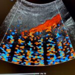
Understanding ultrasound artifacts is essential for accurate interpretation of images. We explain common artifacts, their causes, and how to minimize their effects for improved diagnostic precision.
Discover the world of ultrasound contrast agents, used to enhance imaging clarity. Learn how these agents improve diagnostic accuracy, and explore their applications in various medical fields.
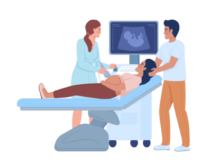
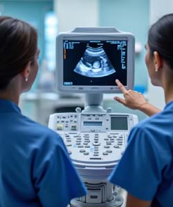
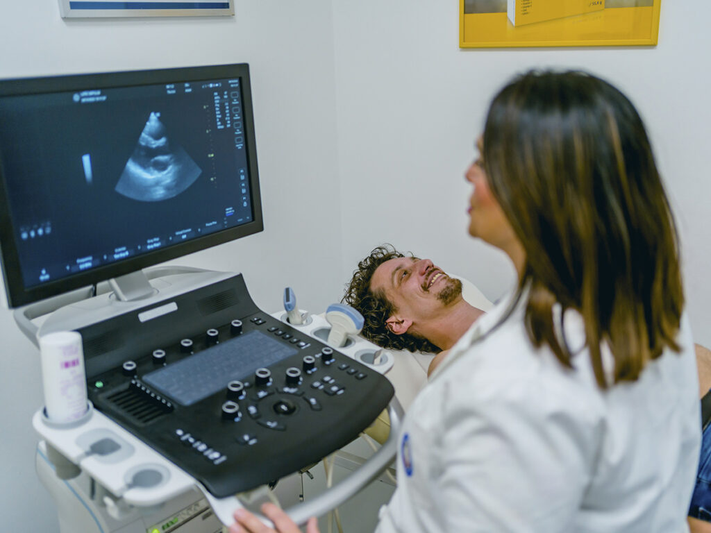
Gain insights into the different ultrasound modes, from B-mode and Doppler imaging to M-mode and 3D/4D ultrasound. These modes are critical for providing detailed views of anatomical structures and blood flow.
We cover a wide range of topics, from ultrasound basics to advanced techniques.
Expert Insights
Our content is backed by radiology professionals, ensuring you receive accurate and relevant information.
Up-to-Date Resources
Stay current with the latest advancements and trends in ultrasound technology and radiology education.
User-Friendly Content
Whether you're a student or a seasoned practitioner, our content is tailored to meet your needs.
PROFESSIONAL REFERENCES
Resources
Radiopaedia – Ultrasound Imaging Library
Radiopaedia is a comprehensive online resource for radiology professionals and students. It offers an extensive library of ultrasound cases, images, and educational articles contributed by radiologists worldwide.
American Institute of Ultrasound in Medicine (AIUM)
The American Institute of Ultrasound in Medicine (AIUM) provides a wealth of educational materials on ultrasound use, including guidelines, tutorials, and webinars. AIUM’s resources cover a broad spectrum of topics.
SonoWorld offers free access to a large collection of ultrasound cases, expert lectures, and technical articles. This resource is especially useful for students and clinicians looking to deepen their understanding of ultrasound applications.
The Society of Radiologists in Ultrasound (SRU) is known for setting the standard in ultrasound use across various medical disciplines. SRU offers guidelines, official reports, and position papers on ultrasound techniques and diagnostic criteria.
US Tip has been my go-to resource for reliable information on ultrasound imaging. Their articles are detailed and accessible, which has helped me stay updated with the latest in radiology.
Mary James
As a radiology student, I found US Tip incredibly helpful. The guides on ultrasound procedures and artifacts have made complex topics easy to understand, and I feel more confident in my studies.
William Scott
I was impressed with the depth of content on ultrasound probes and transducers. US Tip explains advanced concepts in a clear, professional way—highly recommended for anyone in the field.
Dr. Alan Williams
US Tip has set a high standard for ultrasound education. The safety guidelines are particularly useful, ensuring I’m always updated on best practices in patient care.
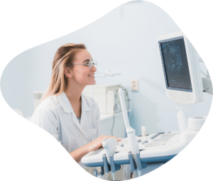
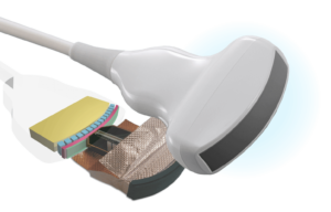
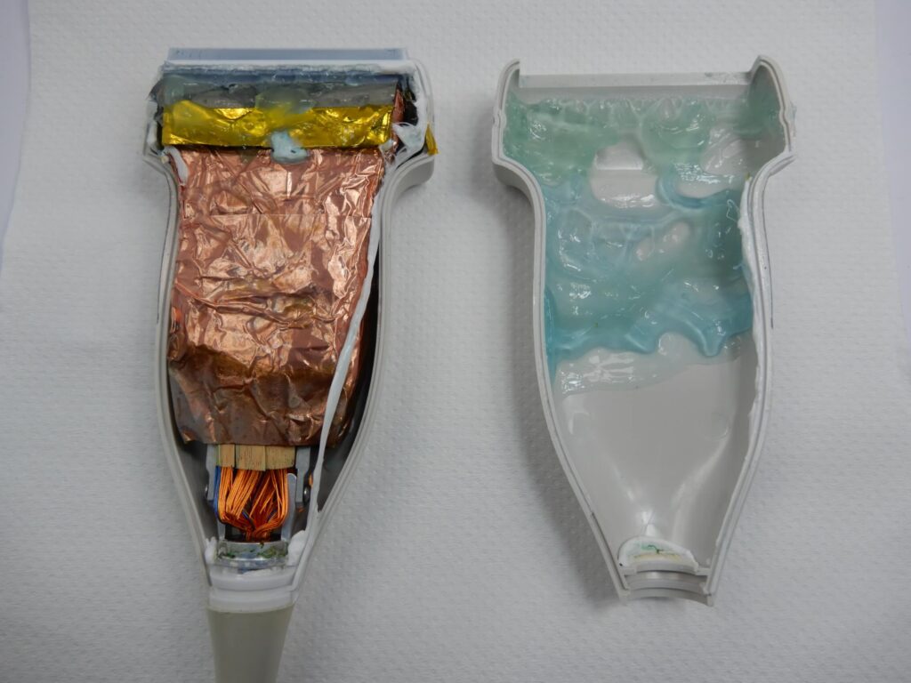
Ultrasound Probes and Transducers
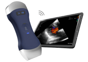
Explore the various types of ultrasound probes and transducers used in medical imaging. Find out which probes are ideal for specific applications, and understand the technology behind these essential tools.
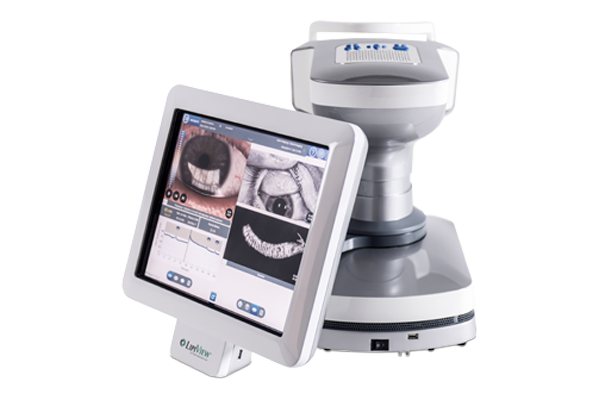
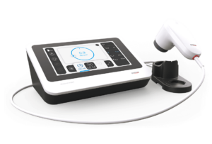
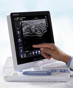
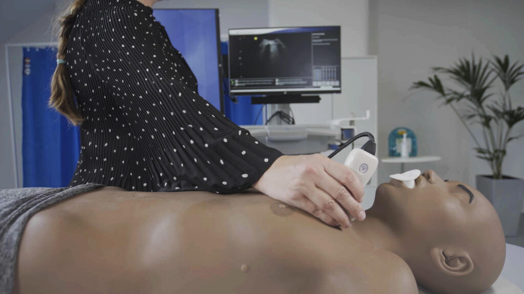
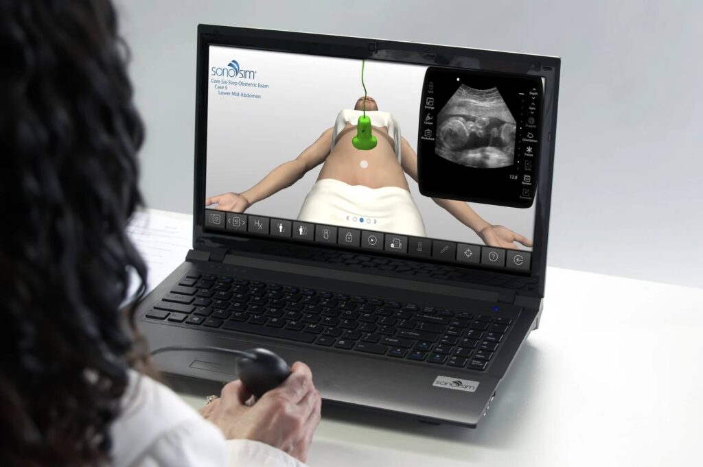
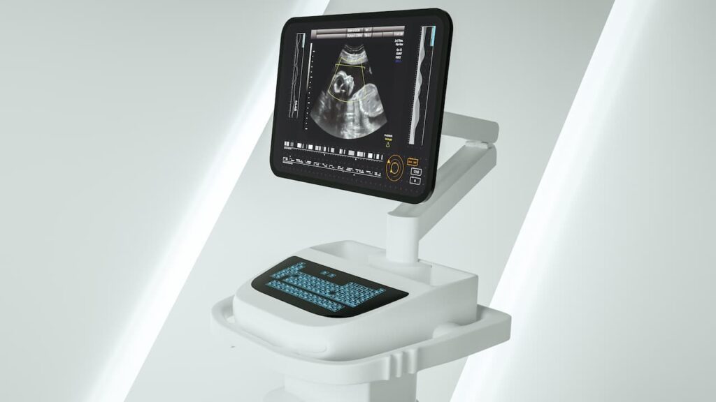
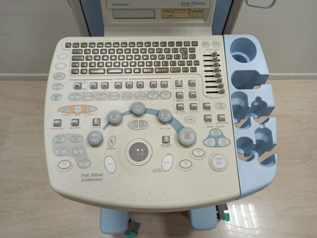
Stay informed about the latest advancements in ultrasound systems. From portable ultrasound machines to state-of-the-art diagnostic systems, we cover the technologies that are shaping the future of medical imaging.
Safety is paramount when using medical ultrasound. We provide guidelines on ultrasound safety to ensure effective and secure usage, including information on thermal and mechanical indices and their impact on patient health.